APPLICATIONS OF TECHNOLOGY:
- Analyzing material interfaces
- Imaging classes of materials that are challenging to image conventionally
ADVANTAGES:
- High signal efficiency
- Good transfer of low spatial frequency information
- Enables imaging while in-focus to minimize signal delocalization
- Anticipated to enable complete aberration correction
- Adaptable to all TEM instruments with pixelated detectors
ABSTRACT:
Berkeley Lab researcher Colin Ophus has developed Matched Illumination and Detector Interferometry-Scanning Transmission Electron Microscopy (MIDI-STEM), a technology for visualizing soft matter, and the interface between hard and soft matter, in-focus, at atomic or near-atomic resolution to enable efficient phase-contrast imaging in STEM. The technology addresses the challenge of imaging the following classes of materials, which are difficult to image conventionally:
- Hard-soft interfaces
- Low-atomic number nanoparticles
- Biomolecules such as 1D and 2D protein structures
- Zeolites and metal-organic frameworks (MOFs)
- 2D materials such as graphene, MoS2, and BN that scatter weakly
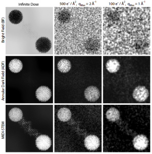
As indicated in Figure 1, a multislice simulation of a DNA snippet connecting two gold nanoparticles – an archetypal hard-soft interface – illustrates the advantages of MIDI-STEM imaging relative to Bright Field (BF)- and Annular Dark Field (ADF)-STEM. MIDI-STEM uses far less electron dose than BF-STEM and ADF-STEM so it can be used on dose-sensitive soft matter, and is adaptable to all TEM instruments with pixelated detector.
Regular electron microscopy methods used by biologists require thousands of identical particles isolated from each other; therefore, many soft matter samples cannot be used. Cryo-electron-microscopy offers in-focus imaging only if the sample is isolated far enough away from other structures so defocused signals do not overlap. 3D reconstruction using Cryo-EM requires thousands of images. In addition, many samples including hard-soft interfaces such as functionalized nanoparticles, cannot be imaged with Cryo-EM.
Figure 1: MIDI-STEM imaging relative to Bright Field (BF)- and Annular Dark Field (ADF)-STEM.
DEVELOPMENT STAGE: Proven principle
STATUS: Patent pending. Available for licensing or collaborative research.
FOR MORE INFORMATION:
Ophus, C., Ciston, J., Pierce, J., Harvey, T. R., Chess, J., McMorran, B. j., Czarnik, C., Rose, H. H., Ercius, P. “Efficient Linear Phase Contrast in Scanning Transmission Electron Microscopy with Matched Illumination and Detector Interferometry.” Publication in press (2015).
