APPLICATIONS:
- Magnetic resonance imaging (MRI) in low field
- Imaging of joints, peripheral regions of the body
- Screening for tumors
- Obtaining direct stereochemical information using J (scalar)coupling spectroscopy in low magnetic field
- Spectroscopy of heterogeneous samples which are subject to spatial variation in magnetic susceptibility, e.g. oil well logging, in vivo spectroscopy, living organisms
- Multinuclear nuclear magnetic resonance (NMR) studies
- NMR spectroscopy of metals
ADVANTAGES:
- Removes major technical barriers to developing lower cost MRI scanners and NMR spectrometers
- Open system
- Uses magnetic fields 10,000 times weaker than conventional MRI
- Lower field homogeneity requirements (only 10,000 ppm to achieve 1 mm resolution)
- Excellent T1-contrast

ABSTRACT:
John Clarke, Alexander Pines, and colleagues have pioneered systems for Nuclear Magnetic Resonance (NMR) and Magnetic Resonance Imaging (MRI) that work in extremely low magnetic fields, typically 1-100 microtesla. They achieved this breakthrough by combining sample prepolarization with ultrasensitive magnetic field detection by Superconducting QUantum Interference Devices (SQUIDs).
Since low magnetic fields are easier and less costly to generate than the high fields necessary for conventional high-field MRI (1.5 tesla), this technique paves the way for developing MRI scanners for certain applications that are less expensive than current commercial systems. Furthermore, the demands on magnetic field homogeneity are greatly reduced at low fields.
The Berkeley Lab researchers achieved narrow (~ 1 Hz) NMR lines from protons in liquids in fields of about 100 microtesla even though the homogeneity was only about 1 part in 10,000. In the context of MRI, the linewidth of the NMR signal determines the spatial resolution of the image for a given strength of magnetic field gradient. The researchers exploit this narrow linewidth to acquire MRIs with 1-mm resolution using a static field of 132 microtesla and field gradients of the order of 100 microtesla/meter three orders of magnitude smaller than the gradients used in conventional high-field MRI. Clarke, Pines, and colleagues prepolarize the sample in a field of 100-300 millitesla. This field is removed before the image is acquired in the much lower field. This technique differs from the standard method in which the protons are polarized and the signal is detected in the same high field. The low-field operation is made possible because a SQUID magnetometer has a magnetic field sensitivity that is independent of frequency. In contrast, the sensitivity of conventional NMR detectors, which rely on Faradays Law, scales linearly with frequency and thus with the magnetic field.
Figure 1 shows two images acquired at 5.6 kHz. Figure 1(a) is a magnetic resonance image of a sliced pepper, while Fig. 1(b) is an MRI slice of an intact pepper obtained by means of a slice-selecting gradient.
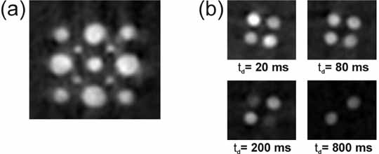
Studies of phantoms with a paramagnetic salt dissolved in water to reduce the longitudinal relaxation time T1 show that excellent T1-contrast images can be obtained see Fig. 2. This technique offers the possibility of low-cost screening for tumors.

STATUS: Issued U. S. Patent #6,885,192.
FOR MORE INFORMATION PLEASE SEE:
McDermott R., Trabesinger A.H., Mück M., Hahn, E., Pines, A., Clarke, J., “Liquid-State NMR and Scalar Couplings in Microtesla Magnetic Fields,” Science 2002, 295, 2247-49
REFERENCE NUMBER: IB-1729
SEE THESE OTHER BERKELEY LAB TECHNOLOGIES IN THIS FIELD:
