APPLICATIONS OF TECHNOLOGY:
- Clinical and experimental MRI
- Portable MRI
- MRI for patients with metal/electronic devices
ADVANTAGES:
- Higher resolution blood flow imaging compared to existing MRI machines
- Measures regional blood flow parameters, not only local velocities
- Images have less background noise
- May be implemented, with minor modifications, in existing MRI machines
ABSTRACT:
Alexander Pines and a team of scientists at Berkeley Lab have invented a technology that enhances conventional MRI to measure fluid flow in microfluidic chips, a model for blood flow in organisms. The invention, which produces high-resolution, time-sequenced images of fluid in channels, could be applied to vessels deep within the body.
Existing MRI machines have a limited ability to image tissue deep in the body with high resolution or to measure blood flow. The Berkeley Lab device overcomes these limitations by encoding a tiny volume of fluid with spatial information when it is in a certain, potentially deep, location. The fluid then naturally passes to a larger, more superficial vessel where a separate, remote detector with a low filling factor receives the encoded information to construct a high-resolution, low-background image of the upstream voxel (a 3-D point denoting a location) and to measure either instantaneous flow at the voxel or average flow from the voxel to the superficial vessel. Clinically, the technology could be applied to non-invasively examine minute parts of the brain affected by stroke or any other organ damaged by insufficient blood flow. It could also be modified to enhance the typically low sensitivity of functional MRI used to monitor levels of activity or metabolism in certain regions of the brain.


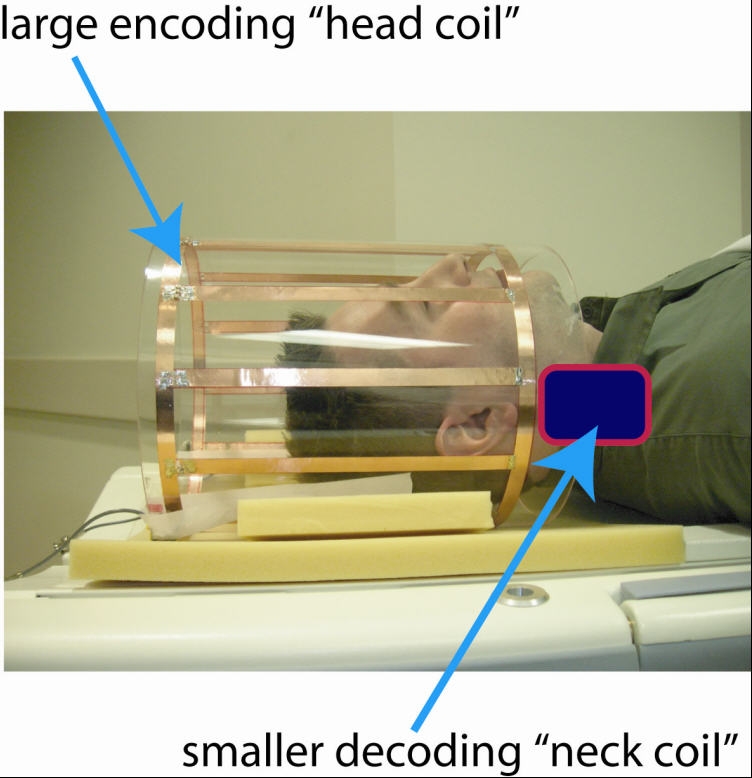
Figure 1. In one potential application of the invention, a head coil is used to spatially encode the spins of atomic nuclei in the blood in the brain. The encoded blood travels to the jugular vein (or any superficial vessel), and a remote neck coil decodes the spatial information to generate an image of the cranial vessels upstream. (hv represents the photon energy from the decoding coil).
The initial polarization and encoding may be done using the powerful fields of a conventional MRI machine to which a small remote detector (approximately 5 cm x 3 cm x 2 cm) can be easily added. The initial polarization and encoding could also be done using weak fields such as those produced by the earth itself or portable magnets and coils mounted on a device that fits around the body part to be imaged. A particularly sensitive remote detector would read these weaker signals. The ability to operate using either powerful or weak fields means that the Berkeley Lab technology potentially circumvents the need for large superconductor magnets to generate the powerful fields used in conventional, immobile MRI machines. This could enable the development of portable, less expensive devices used for patients who are obese, claustrophobic or living in remote regions. The absence of powerful magnets would enable patients with metal implants or electronic monitoring devices to get MRI.


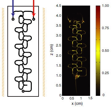
Figure 2. The first panel shows a schematic of the microfluidic chip. Panels A and B show MR images with ultrahigh time resolution demonstrating liquid benzene (red) and acetonitrile (blue and yellow) flowing through the channels of the chip. The remote detector was placed at the outflow of the chip, not directly over the channels shown.
DEVELOPMENT STATUS: Modeled concept. Additional R&D required to develop a prototype.
STATUS: Issued US Patent 8570035 available at www.uspto.gov. Available for licensing or collaborative research.
FOR MORE INFORMATION:
SEE THESE OTHER BERKELEY LAB TECHNOLOGIES IN THIS FIELD:
Optical Atomic Magnetometer for Portable, Inexpensive MRI and Magnetic Microparticle Detection, IB-2232
Portable, High Resolution NMR/MRI in Inhomogeneous Fields, IB-2185
Novel Use of NMR with Microfluidic Lab-on-a-Chip Devices, IB-2212
Low-Field NMR/MRI Using Rotating Frame Gradient Fields, IB-2288
REFERENCE NUMBER: IB-2444