APPLICATIONS OF TECHNOLOGY:
- Medical imaging: MRI, X-ray, photoacoustic tomography (PAT)
ADVANTAGES:
- Visible on multiple, complementary imaging modalities
- Nontoxic, chemically stable contrast agents
- Versatile fabrication using different materials
ABSTRACT:
Fanqing Frank Chen and Louis Bouchard of Berkeley Lab have invented a method for fabricating layered nanoparticles to be used as contrast agents in complementary medical imaging tools such as MRI, X-ray, and photoacoustic tomography (PAT). The scientists produced particles of cobalt (Co) coated with gold (Au) that were stable, biocompatible, and detectable at picomolar (pM) concentrations—107 to 108 times lower than concentrations needed to visualize existing contrast agents such as gadolinium (Gd).
The Co-based, sub-100-nm particles have an unusually high T2 relaxivity of 1 x 10⁷ s-1·mM-1, making them visible on MRI and PAT at concentrations as low as 2.5 pM in agarose phantoms and 50 pM in animal tissue. For PAT, the size and shape of the particles can be altered to tune their peak optical absorption to the incident light, heightening their contrast against surrounding tissue. While MRI clearly shows the entire volume of tissue types, PAT is better for high resolution imaging of tissue margins. Therefore, these nanoparticles provide a single probe visible in both modalities and are ideal for MRI/PAT combinations that offer a comprehensive picture.
One of the most important challenges in imaging technology, especially MRI, is in developing nontoxic contrast agents that have optimal half-lives; are not oxidized to undetectable states; and are visible at the extremely low concentrations administered in living tissue. The Berkeley Lab invention meets these requirements with an optimal particle size and material composition. It also opens the door for linking biomolecules, such as therapeutic agents or diagnostic markers, to the particle coating.

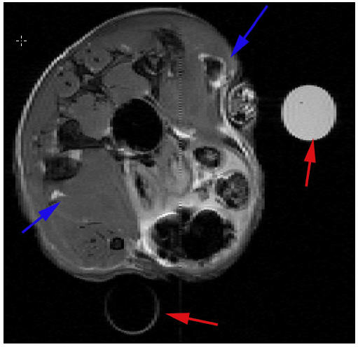
Figure 1. Transverse (axial) MRI of a mouse leg showing muscles injected with Co nanoparticles in phosphate buffered saline (PBS) (blue arrow, upper right) and PBS control (blue arrow, lower left). The red arrows indicate two tubes of water used for MRI slice alignment. MRI parameters: TE = 50 ms; TR = 1 s; field of view, 2.6 x 2.6 cm; slice thickness, 0.5 mm.

Figure 2. Photoacoustic tomography (PAT) imaging of a rat tail joint taken (a) before and (b) after the administration of the nanoparticle contrast agent at 100 pM. In comparison, (c) is a histological photograph of a similar cross section showing the periosteum. In (a) and (b), grayscale is in arbitrary units of relative optical absorption, and xy scales are 1 cm X 1 cm.
DEVELOPMENT STAGE: Proven principle.
STATUS: Published Patent Application WO2010096828 available at www.wipo.int. Available for licensing or collaborative research.
SEE THESE OTHER BERKELEY LAB TECHNOLOGIES IN THIS FIELD:
New Multimodal Probes Compatible with Fluorescence and MRI, JIB-2075
REFERENCE NUMBER: IB-2698